![]() Figure 1 of
Tsai, Mol Vis 2005;
11:71-75.
Figure 1 of
Tsai, Mol Vis 2005;
11:71-75.
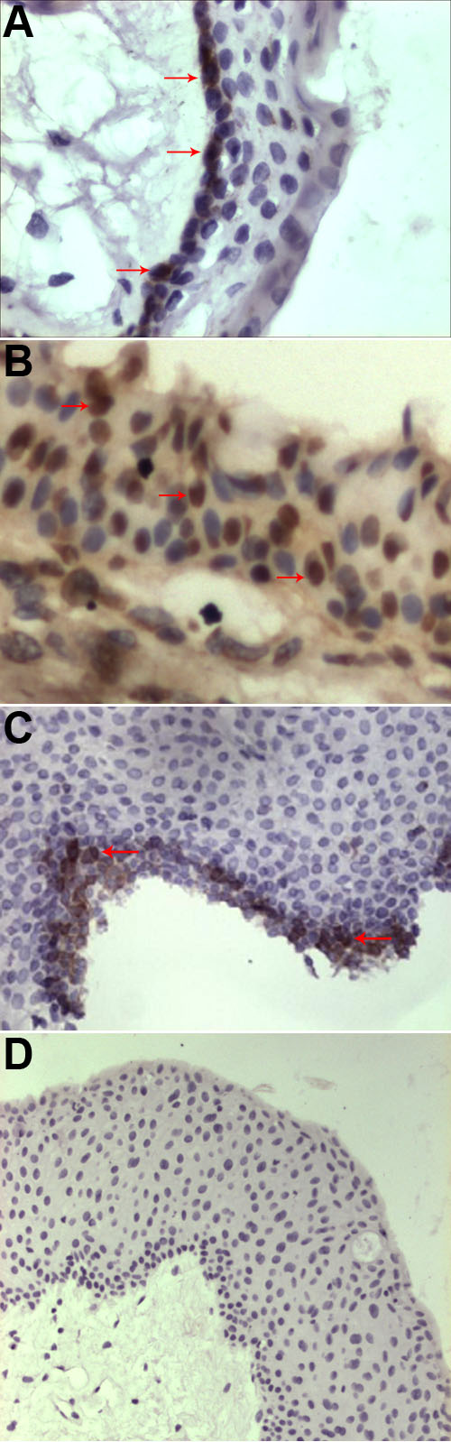
Figure 1. 8-OHdG staining
8-OHdG positive staining is shown in the nucleus of the epithelium, and distributed in the basal (A,B), middle (B), and superficial (B,C) layers of the epithelium. Arrows indicate the immunostained nuclei. No substantial staining is visible in the subepithelial fibrovascular layer or in normal control (D).