![]() Figure 9 of
Vasireddy, Mol Vis 2005;
11:665-676.
Figure 9 of
Vasireddy, Mol Vis 2005;
11:665-676.
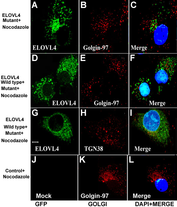
Figure 9. ELOVL4 aggresomes are distinct from Golgi
Cells that express the GFP-Emut (A-C), GFP-Ewt and GFP-Emut (D-I), control (J-L) were treated with the microtubule disrupting drug nocodazole. Treatment with nocodazole disrupted both Golgi and ELOVL4 aggresomes. Fluorescent signals from ELOVL4, golgin-97, and TGN-38 were found to be distributed as spots in the cytoplasm in cells expressing GFP-Emut (A-C) and GFP-Ewt and GFP-Emut (D-I). The ELOVL4 signal did not colocalize with either golgin-97 or TGN-38 indicating that the ELOVL4 aggresomes are distinct from Golgi. Golgin-97 and TGN38 staining is shown in red. Panels C, F, I, and L are the overlays of GFP and Golgi images with DAPI stained nuclei. The scale bar represents 5 μm.