![]() Figure 7 of
Vasireddy, Mol Vis 2005;
11:665-676.
Figure 7 of
Vasireddy, Mol Vis 2005;
11:665-676.
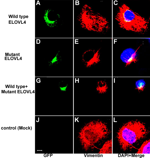
Figure 7. Reorganization of vimentin in the cytoskeleton
COS-7 cells transfected with GFP-Ewt (A-C), GFP-Emut (D-F), GFP-Ewt and GFP-Emut (G-I), and mock transfected control (J-L) were labeled with anti-vimentin antibody. Formation of aggresomes is associated with the reorganization of intermediary filament protein, vimetin.Vimentin is redistributed from the normal reticular pattern into a condensed pattern near the aggregated ELOVL4 protein. Vimentin distribution is shown in B, E, H, and K (red). Nuclei were stained with DAPI. Panels C, F, I, and L are the overlays of first two columns. The scale bar represents 5 μm.