![]() Figure 6 of
Vasireddy, Mol Vis 2005;
11:665-676.
Figure 6 of
Vasireddy, Mol Vis 2005;
11:665-676.
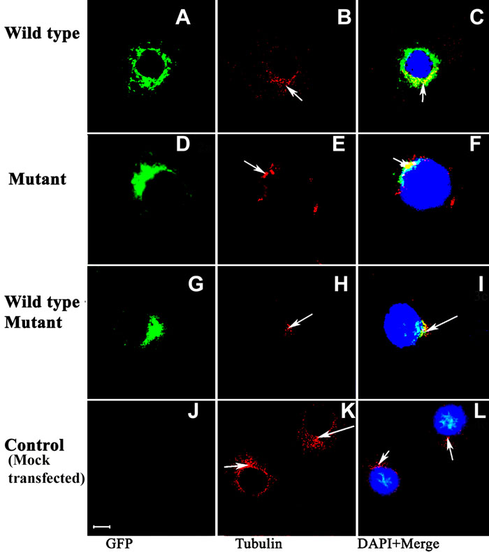
Figure 6. Formation of aggresomes at the MTOC
COS-7 cells were transfected with GFP-Ewt (A-C), GFP-Emut (D-F), GFP-Ewt and GFP-Emut (G-I), and mock transfected with transfection reagents alone (K) and were immunostained with antibodies to the centrosome marker, γ-tubulin. Staining of γ-tubulin is indicated with arrows (red). Overlays of images from first two columns on DAPI-stained nuclei of the respective cells (blue) were shown in panels C, F, I, and L. Physically interacting wild-type and mutant ELOVL4 formed aggresomes near MTOC (microtubule organizing center). The scale bar represents 5 μm.