![]() Figure 2 of
Karan, Mol Vis 2005;
11:657-664.
Figure 2 of
Karan, Mol Vis 2005;
11:657-664.
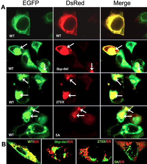
Figure 2. Localization of transfected ELOVL4 fusion proteins
Confocal images were recorded 24 h after transfection. A: wtEGFP/ELOVL4 was cotransfected (1:1) with wtDsRed/ELOVL4 or mutant DsRed/ELOVL4 constructs as indicated in each panel. The green color represents EGFP fused ELOVL4 wild-type protein, and the red color identifies the wild-type or mutant DsRed fused ELOVL4 proteins. When wild-type and mutant proteins were co-expressed, the overlapping (yellow) fluorescence signals were associated with aggregates (right panels). Note the clumped signals (both EGFP and DsRed) in the cells co-expressing mutant ELOVL4 protein (arrows). B: To visualize mislocalized ELOVL4 proteins, recombinant wtEGFP/ELOVL4 and mutant EGFP/ELOVL4 constructs were cotransfected with the ER specific marker, pDsRed2-ER. Mutant ELOVL4 protein signals (green) did not overlap with the ER protein marker signal (red).