![]() Figure 6 of
Yamagami, Mol Vis 2005;
11:632-640.
Figure 6 of
Yamagami, Mol Vis 2005;
11:632-640.
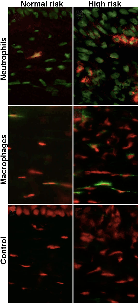
Figure 6. Neutrophil and macrophage infiltration into corneal allografts on day 6
Representative photomicrograph of corneas harvested from normal risk (NR) and high risk (HR) transplant recipients 6 days post-keratoplasty. Representative photographs using anti-Gr-1, anti-F4/80, and nonimmunized control rat IgG2a antibodies were shown. Increased infiltration of neutrophils (Gr-1; red) is observed into the HR grafts compared to the NR grafts. The number of macrophages (F4/80; green) in the HR grafts is high compared with the NR grafts. No positive staining in the NR grafts and the HR grafts were observed with FITC-conjugated rat IgG2a control antibody. SYBR Green (green; top two panels) and PI (red; lower 4 panels) were used for nuclear staining. No positive staining was observed with FITC-conjugated hamster and rat IgG2b control antibody (not shown).