![]() Figure 5 of
Yamagami, Mol Vis 2005;
11:632-640.
Figure 5 of
Yamagami, Mol Vis 2005;
11:632-640.
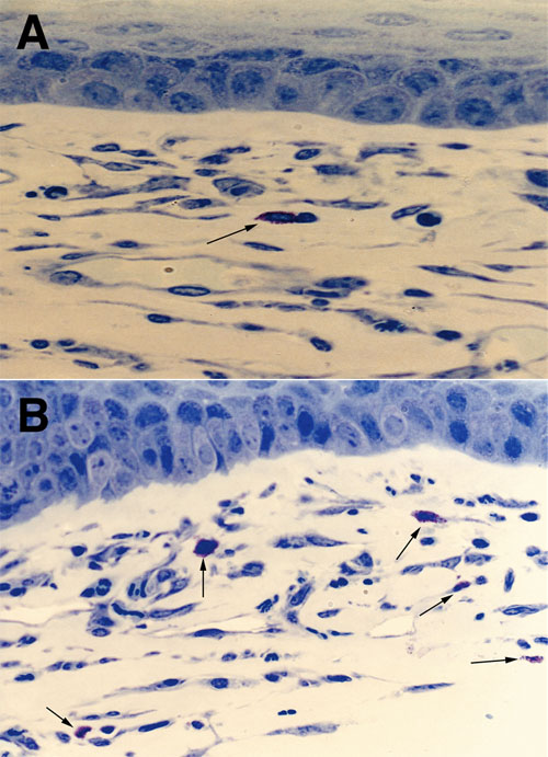
Figure 5. Enhanced presence of mast cells in high-risk transplantation
Representative photomicrograph of corneas harvested from normal-risk (NR; A) and high-risk (HR; B) transplant recipients 6 days post-keratoplasty. Toluidine blue staining of the sections reveals mast cells with characteristic magenta staining metachromatic granules (arrows). Increased infiltration of mast cells is observed into HR grafts (B) compared to NR grafts (A). Ungrafted normal corneas had no mast cells (not shown).