![]() Figure 3 of
Yamagami, Mol Vis 2005;
11:632-640.
Figure 3 of
Yamagami, Mol Vis 2005;
11:632-640.
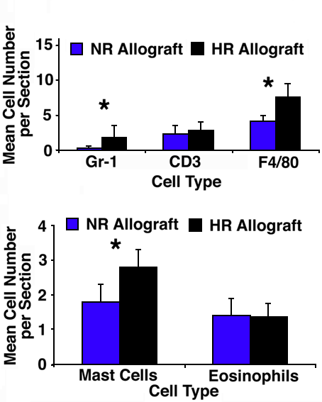
Figure 3. Leukocyte infiltration into corneal allografts on day 3
Allogeneic (C57BL/6) corneal grafts were performed in normal-risk (NR) and high-risk (HR) BALB/c host beds at day 0. At day 3, eyes were harvested and the leukocytic infiltration quantified and subtyped based on immunohistochemical staining with specific membrane markers: anti-Mac-3 for macrophages, anti-CD3 for T cells, and anti-Gr-1 for neutrophils, and histochemical analyses for mast cells and eosinophils. Significantly increased infiltration of neutrophils and macrophages (asterisk indicates p<0.05) were observed into the HR grafts compared to the NR grafts. The error bars represent standard deviation.