![]() Figure 8 of
Scheef, Mol Vis 2005;
11:613-624.
Figure 8 of
Scheef, Mol Vis 2005;
11:613-624.
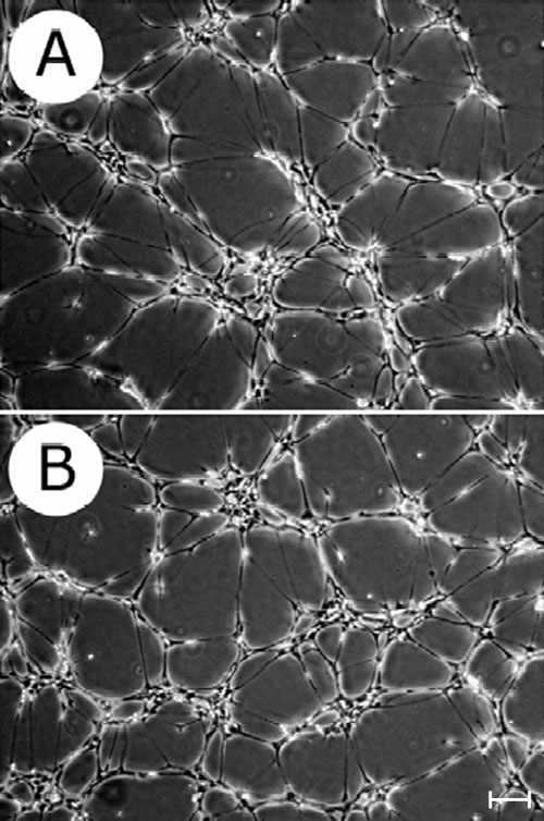
Figure 8. Organization of retinal astrocytes in Matrigel
RAC from wild-type (A) and TSP1-/- (B) cells were plated in Matrigel as described in Methods. The organization of cells into a network was digitally photographed using a phase-contrast microscope after incubation at 33 °C for 10 h. Both cell types exhibited a similar ability to organize into a network in Matrigel. These experiments were repeated three times with similar results. The scale bar represents 40 μm.