![]() Figure 1 of
Scheef, Mol Vis 2005;
11:613-624.
Figure 1 of
Scheef, Mol Vis 2005;
11:613-624.
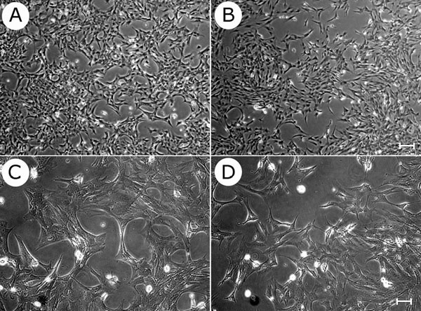
Figure 1. Morphology of mouse retinal astrocytes in culture
Retinal astrocytes in culture (RAC) from wild-type (A,C) and TSP1-/- (B,D) mice were cultured on gelatin coated plates. Cells were photographed using a Nikon phase microscope in a digital format (top 2 panels, 40x; bottom 2 panels, 100x). RAC from wild-type and TSP1-/- mice share a similar morphology. The scale bar represents 40 μm in A,B and 100 μm in C,D.