![]() Figure 3 of
Jablonski, Mol Vis 2005;
11:569-581.
Figure 3 of
Jablonski, Mol Vis 2005;
11:569-581.
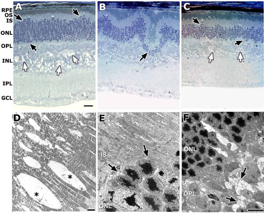
Figure 3. Detailed structural analysis
Light-level histology and electron microscopic images of the retinas of male 44TNJ mutant mice at various ages are presented in this figure. Representative images of retinal structure from 2 week (A), 14 week (B), and 38 week (C) 44TNJ mutant mice are shown. Displacement of photoreceptor nuclei (black arrows) and schisis of the inner nuclear layer (INL; white arrows) are present as early as 2 weeks (A). The same morphological features are present with no progression until 38 weeks (C). Less frequently, large clusters of displaced nuclei appear to have migrated into the INL (arrow in B). D-F: Representative electron micrographs taken from the 14 week old 44TNJ mouse are shown. D: Gaps are present between outer segments (OS) with occasional debris (asterisks). E: In areas where the photoreceptor nuclei are displaced into the inner segments (IS), the adherens junctions that comprise the outer limiting membrane are absent (arrows). F: The outer plexiform layer (OPL) structure is disrupted and filled with debris (arrows). The retinal pigment epithelium (RPE), outer nuclear layer (ONL), inner plexiform layer (IPL), and ganglion cell layer (GCL) are also identified. Scale bars represent 20 μm in A-C and 2 μm in D-F.