![]() Figure 2 of
Jablonski, Mol Vis 2005;
11:569-581.
Figure 2 of
Jablonski, Mol Vis 2005;
11:569-581.
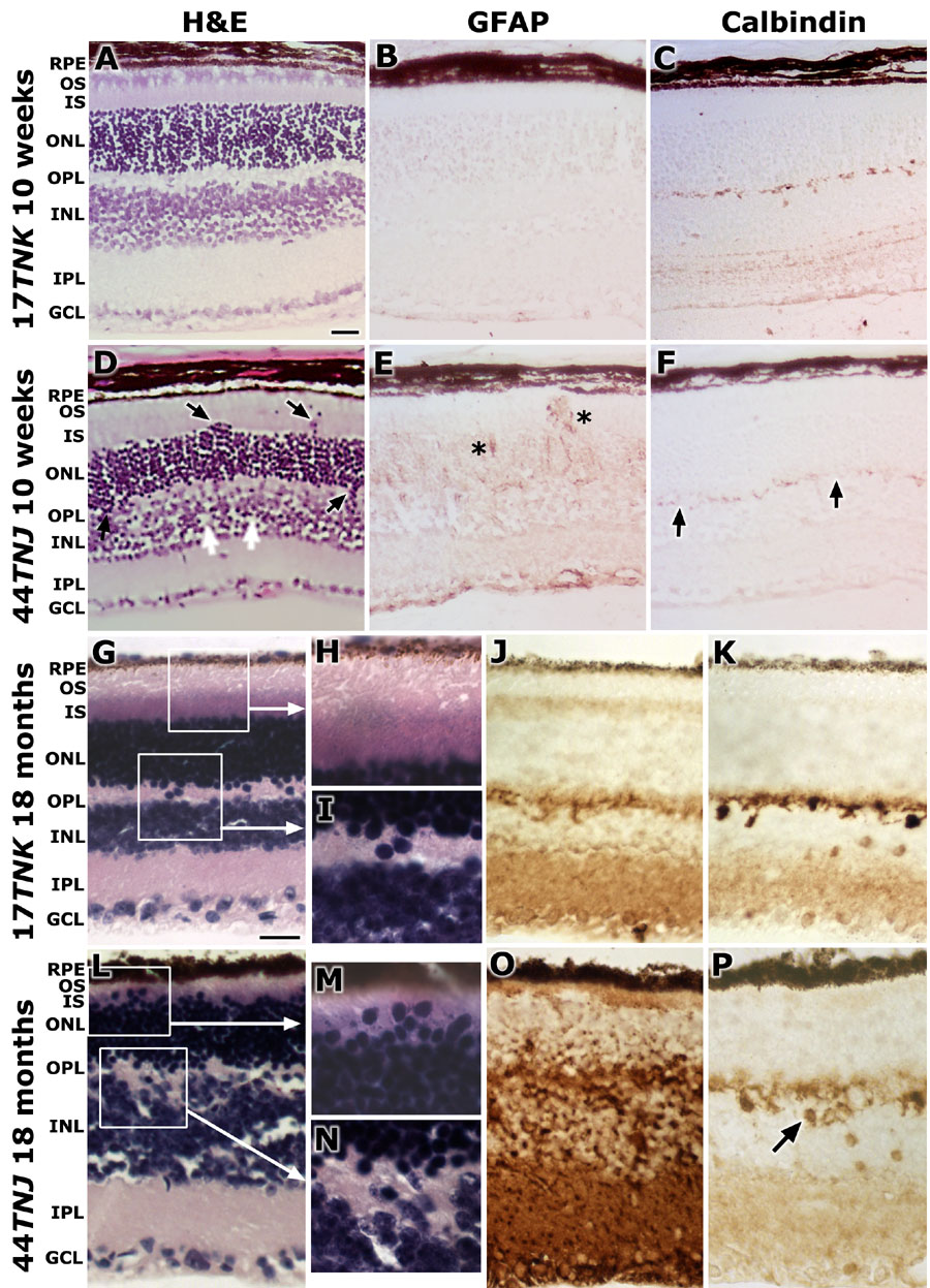
Figure 2. Histological and immunohistochemical analyses of retinas from control and 44TNJ mice at 10 weeks and 18 months
Representative images of retinal structure (A) and immunohistochemical localization of GFAP (B) and calbindin (C) from 10 week old 17TNK control mice are shown; while retinal sections from 10 week old 44TNJ mutant mice are illustrated in D, E, and F, respectively. Note the displacement of photoreceptor nuclei (black arrows) and the focal splitting of the INL (white arrows) in retinas from 44TNJ mice (D). GFAP immunolabeling was elevated in the retinas of mutant mice (asterisks in E). In contrast, calbindin immunolabeling was decreased in those same mice (black arrows in F). Similar representative images from 18 month old mice are illustrated in G-P. With aging, there is no change in retinal structure in the 17TNK control mice (G-I). However, the intensity of the GFAP (J) and calbindin (K) immunolabeling increased slightly. In the retinas from 18 month old 44TNJ mice, there was a shortening of photoreceptor inner segments (IS) and outer segments (OS; L,M) along with further displacement of nuclei into the areas of IS, OS and the outer plexiform layer (OPL; L,N). Moreover, the inner nuclear layer (INL) appeared to split, thus allowing the OPL to fill the gaps (L,N). GFAP levels were greatly increased (O) over the age-matched controls. Calbindin labeling was reduced in intensity and many calbindin-positive horizontal cells were localized deeper into the INL (black arrow in P). The retinal pigment epithelium (RPE), outer nuclear layer (ONL), inner plexiform layer (IPL), and ganglion cell layer (GCL) are also identified. Scale bars represent 10 μm.