![]() Figure 1 of
Jablonski, Mol Vis 2005;
11:569-581.
Figure 1 of
Jablonski, Mol Vis 2005;
11:569-581.
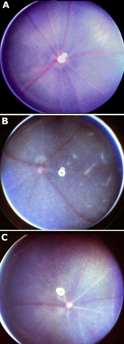
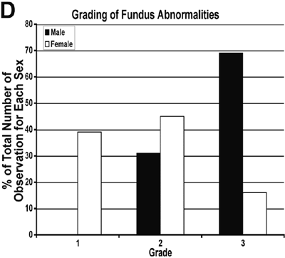
Figure 1. Fundus images and grading of fundus abnormalities
Representative fundus images from a male 17TNK control mouse (A), a male 44TNJ mouse with both intraretinal flecks and streaks throughout the posterior pole (B), and a female 44TNJ mouse with only intraretinal flecks (C) are shown. D: A histogram of fundus grade using our scale (grade 1: normal fundus; grade 2: fundus that presents with small focal intraretinal microflecks; grade 3: fundus that presents with larger streak-like flecks in addition to intraretinal microflecks) shows that 100% of the male G4 44TNJ mice have fundus abnormalities, while only 61% of the female mice from G4 present with fundus flecks or streaks.