![]() Figure 7 of
Kumar, Mol Vis 2005;
11:561-568.
Figure 7 of
Kumar, Mol Vis 2005;
11:561-568.
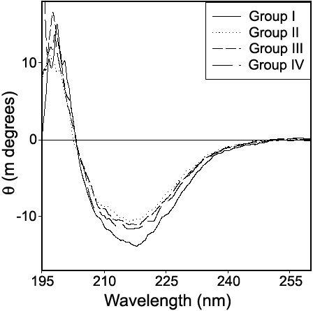
Figure 7. Secondary structure of αL-crystallin
Secondary structure of αL-crystallin from different groups was assessed by far UV CD spectroscopy using 0.10 mg/ml protein in 20 mM sodium phosphate buffer (pH 7.2).