![]() Figure 5 of
van der Spuy, Mol Vis 2005;
11:542-553.
Figure 5 of
van der Spuy, Mol Vis 2005;
11:542-553.
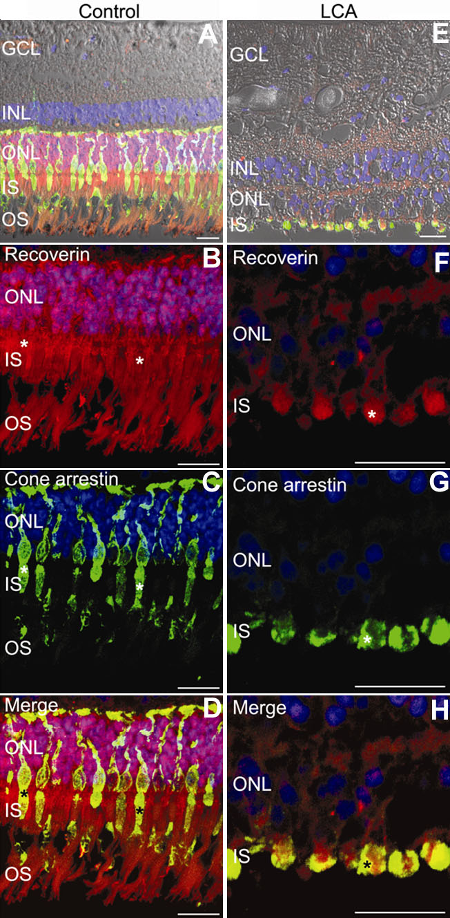
Figure 5. Labeling of the control and LCA retina with Recoverin N-16 (rod and cone recoverin) and LUMIf (cone arrestin)
Double immunofluorescent confocal microscopy of the control, and LCA retina labeled with antibodies for rod and cone recoverin (N-16, red), and cone arrestin (LUMIf, green). The retinal nuclei were counterstained with DAPI (blue). The control (A) and LCA (E) retinal sections were visualized with bright field and differential interference contrast (Nomarski) optics, and the immunofluorescent confocal Z sections and bright field images were overlaid (A,E). The control (B-D), and LCA (F-H) sections were also visualized at higher magnification without Nomarski optics. The control and LCA sections were labeled with antibodies for recoverin shown in red (B,F), cone arrestin shown in green (C,G), and colocalization of the recoverin and cone arrestin label shown in yellow (asterisks), were visualized by merging the images of the confocal Z sections (D,H). The photoreceptor outer segments (OS), photoreceptor inner segments (IS), outer nuclear layer (ONL), inner nuclear layer (INL), and ganglion cell layer (GCL) are labeled. The scale bars represents 20 μm.