![]() Figure 2 of
van der Spuy, Mol Vis 2005;
11:542-553.
Figure 2 of
van der Spuy, Mol Vis 2005;
11:542-553.
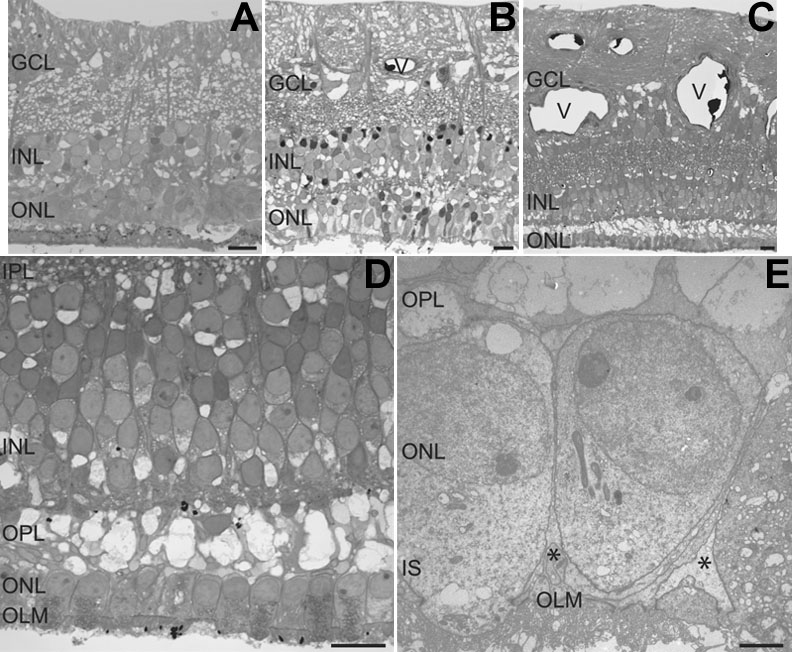
Figure 2. Morphology of the LCA retina examined by light and electron microscopy
Light microscopy of semithin sections (1 μm) from peripheral (A), midperipheral (B), and macular (C,D) areas of the LCA retina. Electron micrograph of an ultrathin section from the macular area of the LCA retina is shown in panel E. The photoreceptor inner segments (IS), outer nuclear layer (ONL), inner nuclear layer (INL), outer limiting membrane (OLM), outer plexiform layer (OPL), inner plexiform layer (IPL), and ganglion cell layer (GCL) are labeled. Blood vessels are identified with the letter "V" in their lumen. Müller cell processes are identified with asterisks ("*"). The scale bars represent 20 μm.