![]() Figure 4 of
Jin, Mol Vis 2005;
11:535-541.
Figure 4 of
Jin, Mol Vis 2005;
11:535-541.
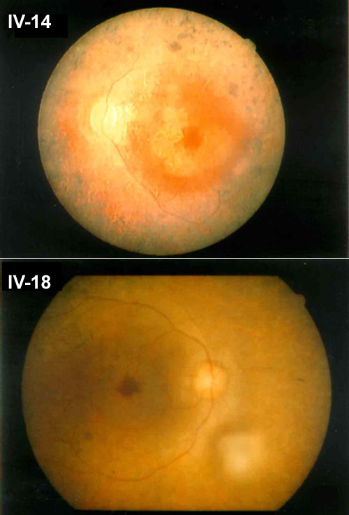
Figure 4. Fundus photographs of two affected patients
Fundus image (top) for the left eye of IV-14 at 51 years showed severe retinal degeneration and moderate pigmentary changes with bone spicule-like pigmentation. Fundus photograph (bottom) from the right eye of IV-18 at 35 years showed mild degeneration and scarcely any pigmentation.