![]() Figure 3 of
Bantseev, Mol Vis 2005;
11:518-523.
Figure 3 of
Bantseev, Mol Vis 2005;
11:518-523.
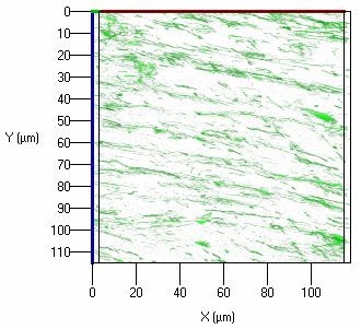
Figure 3. Mitochondrial morphology and distribution in the superficial cortical fiber cells
In the lens superficial cortical fiber cells, the mitochondria are not as dense as that of the epithelial cells; rather, they are more elongated and aligned along the long axis of the fiber cells. Images of the superficial cortex were obtained 10-15 μm below lens epithelium. This three dimensional animation was reconstructed from 18 z-stacks showing the distribution and morphology of the mitochondria in the superficial cortical fiber cells. The z-series were taken 8-10 μm below the lens surface (immediately below the lens epithelial cells) at an increments of 10 μm for up to 170 μm.
Note that the slide bar at the bottom of the quicktime movie can be used to manually control the flow of the movie. If you are unable to view the movie, a representative frame is included below.
| This animation requires Quicktime 6 or later. Quicktime is available as a free download. |