![]() Figure 2 of
Bantseev, Mol Vis 2005;
11:518-523.
Figure 2 of
Bantseev, Mol Vis 2005;
11:518-523.
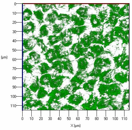
Figure 2. Mitochondrial morphology and distribution in the lens epithelium
Numerous mitochondria arranged radially about the nucleus are seen in the lens epithelium. Shorter and not as dense mitochondria are seen in the cell periphery. This three dimensional animation was reconstructed from 25 z-stacks of the entire thickness of lens epithelium. The bar is equal to 10 μm.
Note that the slide bar at the bottom of the quicktime movie can be used to manually control the flow of the movie. If you are unable to view the movie, a representative frame is included below.
| This animation requires Quicktime 6 or later. Quicktime is available as a free download. |