![]() Figure 1 of
Bantseev, Mol Vis 2005;
11:518-523.
Figure 1 of
Bantseev, Mol Vis 2005;
11:518-523.
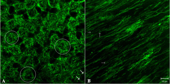
Figure 1. Distribution of the mitochondria in lens epithelial and superficial cortical fiber cells
Whole bovine lenses were loaded with the mitochondria specific dye tetramethylrhodamine ethyl ester (TMRE) and images at the lens equator were obtained as described in Methods. Very different morphology and distribution patterns were seen between the epithelial and superficial cortical fiber cells. In lens epithelial cells (A) the mitochondria exhibit extraordinary diversity with all gradations in shape and size, from distinct minute granules seen in the cell periphery to centrally located numerous threads that are up to 10 μm long (two white arrows point to one example). Those numerous threads, loops and rings appear to form a network arranged radially about the nucleus of the cell. This elaborate network may involve many of the mitochondria or only a few of them. In the lens superficial cortex (B) the mitochondria are not as dense, more elongated (up to 65 μm long), distinctly separated, occasionally branched (the branching is indicated by white arrows) and with no particular association with the nuclei. Images of the superficial cortex were obtained 10-15 μm below the lens epithelium. The bar represents 10 μm.