![]() Figure 6 of
Mullins, Mol Vis 2005;
11:509-517.
Figure 6 of
Mullins, Mol Vis 2005;
11:509-517.
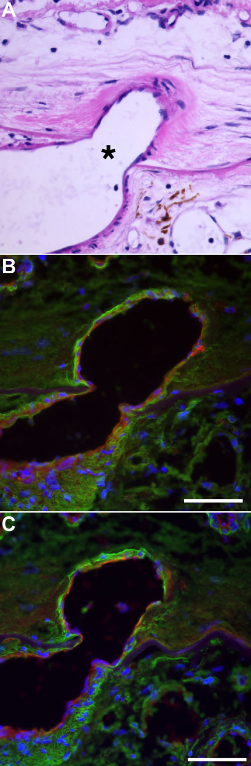
Figure 6. Histochemistry of a feeder vessel in a CNVM
A: Hematoxylin and eosin stain of a large vessel breaching Bruch's membrane (at the asterisk). B: Colocalization of collagen type IV (green) and SBA (red) in this vessel. Note the reactivity of SBA in the endothelium and surrounding matrix. C: Colocalization of collagen type IV (green) and sWGA (red) in the same vessel shown in A and B. Note that, unlike SBA, most of the sWGA binding is present on the endothelium. All scale bars represent 50 μm.