![]() Figure 5 of
Mullins, Mol Vis 2005;
11:509-517.
Figure 5 of
Mullins, Mol Vis 2005;
11:509-517.
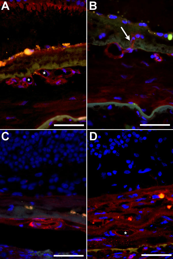
Figure 5. Lectin reactivity of vessels in choroidal neovascular membranes
A: VVA labeling of cone inner segments and CNV vasculature (Case 2). B: An SBA-reactive vessel in a CNVM sends a branch (arrow) into the layer of BlamD (Case 2). C: A flat layer of vessels in Case 1 is positive for SBA. D: Vessels in Case 1 reactive for sWGA (asterisk). Red fluorescence indicates lectin labeling, blue fluorescence represents DAPI labeling, and orange-yellow fluorescence indicates RPE lipofuscin autofluorescence. The scale bars represent 50 μm.