![]() Figure 4 of
Mullins, Mol Vis 2005;
11:509-517.
Figure 4 of
Mullins, Mol Vis 2005;
11:509-517.
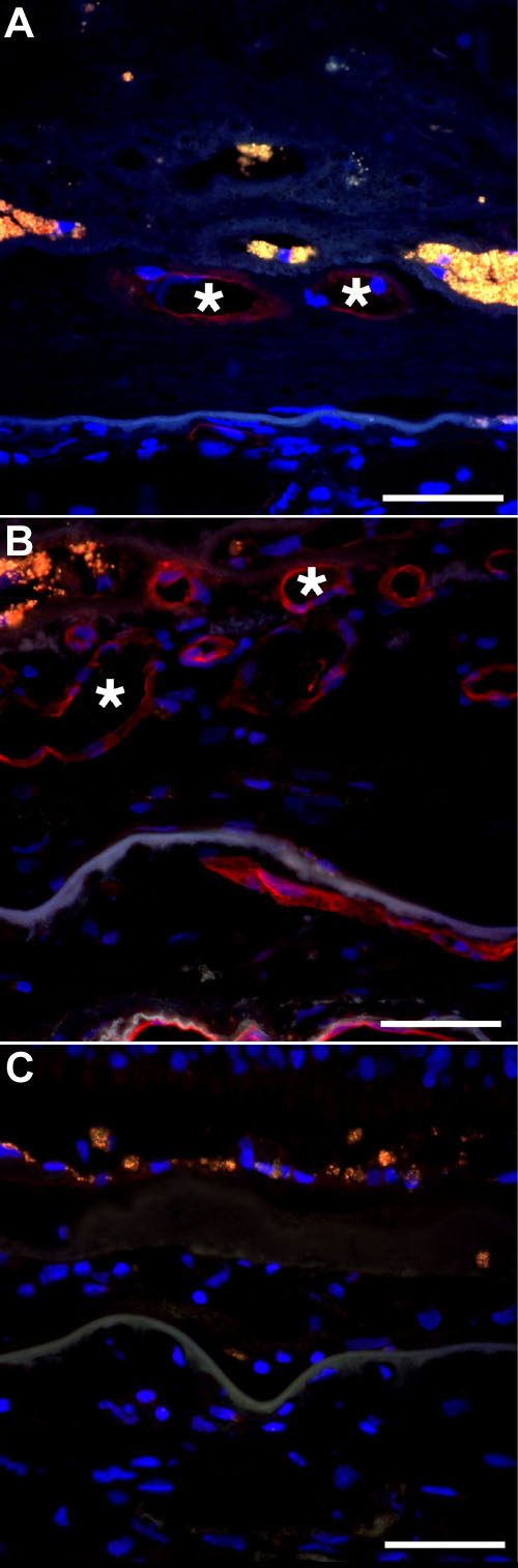
Figure 4. Reaction of choroidal neovessels with the fucose-binding lectin UEA-I
All viable vessels in Case 1 (A) and Case 2 (B) were labeled with UEA-I (asterisks). C: Very minor fluorescence of some vessels was observed with the avidin-Texas red reagent alone (Case 2). The scale bars represent 50 μm. Red fluorescence indicates lectin labeling, blue fluorescence indicates DAPI labeling, and orange-yellow fluorescence indicates RPE lipofuscin autofluorescence.