![]() Figure 3 of
Mullins, Mol Vis 2005;
11:509-517.
Figure 3 of
Mullins, Mol Vis 2005;
11:509-517.
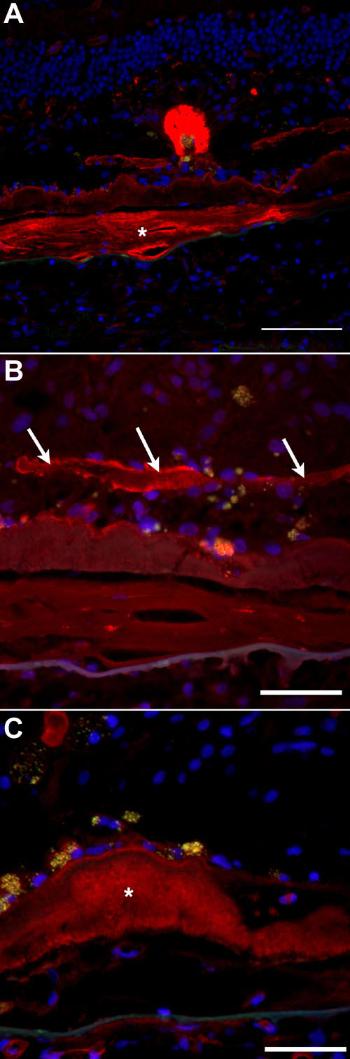
Figure 3. Matrix labeling with some lectins
A: PNA labels a photoreceptor rosette (previously described [26]) and the material within the scar (asterisk). B: VVA reacts with basement membrane material in the subretinal space (arrows). C: When utilized at higher concentrations, sWGA reacts with the layer of basal laminar deposit (asterisk) and choriocapillaris blood vessels. Red fluorescence indicates lectin labeling, blue fluorescence indicates DAPI labeling, and orange-yellow fluorescence indicates RPE lipofuscin autofluorescence. The scale bars represent 100 μm in A and 50 μm in B,C.