![]() Figure 2 of
Mullins, Mol Vis 2005;
11:509-517.
Figure 2 of
Mullins, Mol Vis 2005;
11:509-517.
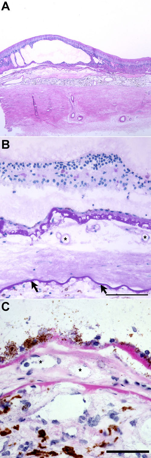
Figure 2. Histopathology of Cases 2 and 3
A: Low magnification view of Case 2 showing cystic changes in the neural retina overlying the area of neovascularization. (H&E stain; collected with a 2.5x objective lens) B: PAS reactivity of BlamD in Case 2 and the outer layers of Bruch's membrane (arrows). Several vascular elements are present in the space between these two matrices (asterisks), the outer nuclear layer is attenuated, and cystic changes are present within the inner nuclear layer (PAS stain). The scale bar represents 100 μm. C: In Case 3, a CNVM is observed between the RPE and the outer layers of Bruch's membrane (H&E stain; asterisks identify blood vessels). The scale bar represents 50 μm.