![]() Figure 1 of
Mullins, Mol Vis 2005;
11:509-517.
Figure 1 of
Mullins, Mol Vis 2005;
11:509-517.
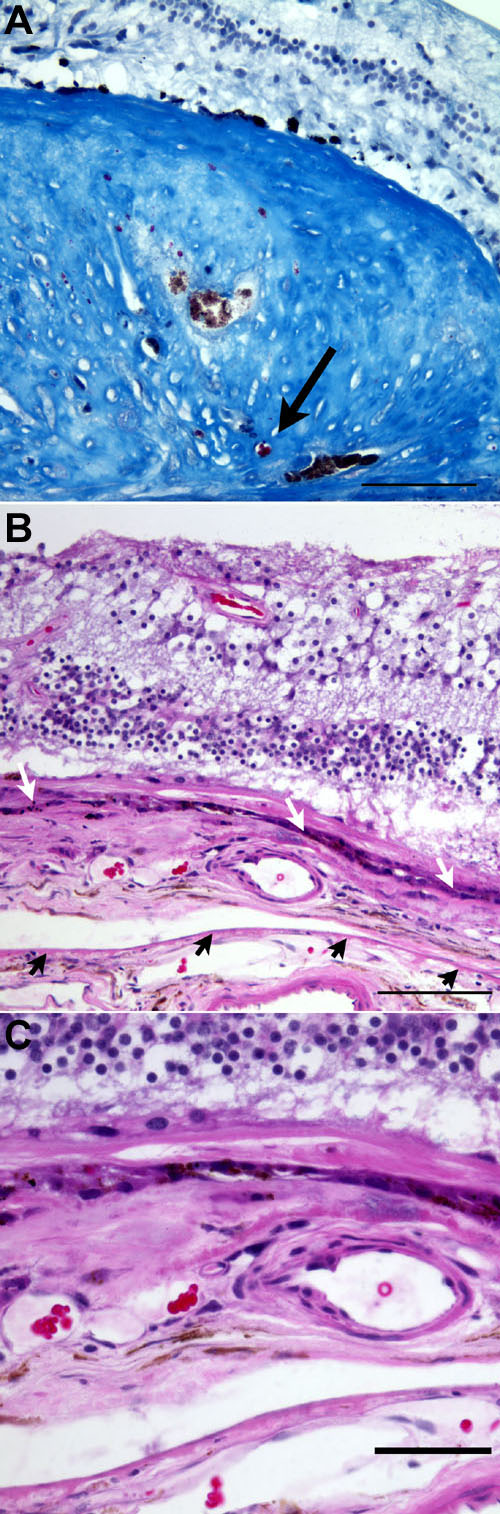
Figure 1. Histopathology of Case 1
A: Area of disciform scarring reacts with Masson's Trichrome to give a blue staining pattern. Note the RPE pigment and the small vessel with red blood cells embedded in the scar (arrow). The outer nuclear layer shows significant degeneration. The scale bar represents 100 μm. B: A subpigment epithelial neovascular membrane is present in this eye, with degeneration of the overlying retina. This membrane is primarily located between the dystrophic RPE (white arrows) and Bruch's membrane (black arrows; H&E stain). The scale bar represents 100 μm. C: Higher magnification of B (H&E stain). The scale bar represents 50 μm.