![]() Figure 6 of
Lewis, Mol Vis 2005;
11:491-500.
Figure 6 of
Lewis, Mol Vis 2005;
11:491-500.
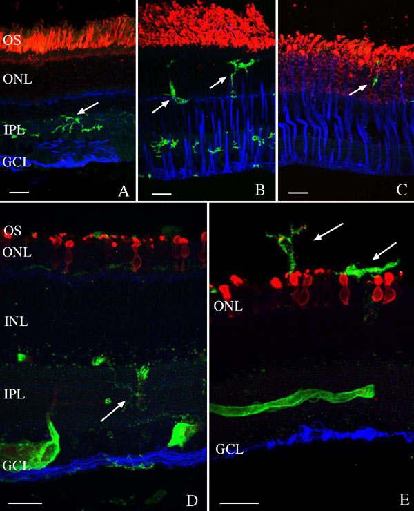
Figure 6. Localization of microglia in normal and detached rabbit and ground squirrel retina
Laser scanning confocal images of rabbit (A,B,C) and California ground squirrel (D,E) retina are shown labeled with the lectin Griffonia simplicifolia (green) and antibodies to rod opsin (red) and glial fibrillary acidic protein (GFAP; blue). A: In normal rabbit retina the lectin labels cells in the inner plexiform layer (IPL), anti-rod opsin labels outer segments, and anti-GFAP labels astrocytes and Müller cells in the inner retina. B: At one day of detachment, lectin-labeled cells are observed in the outer nuclear layer (ONL) as the outer segments (OS) begin to degenerate. C: At 7 days of detachment, few lectin-labeled cells are observed, rod photoreceptors are highly degenerate and are labeled with anti-opsin throughout their plasma membrane, and anti-GFAP is elevated in Müller cells. D,E: Two examples of ground squirrel retina detached for 7 days. Few lectin-labeled cells (arrows) are observed in either the retina or the subretinal space. Anti-opsin labeling illustrates the highly degenerate morphology of the photoreceptors, labeling the truncated OS and the entire plasma membranes of the cells. No increase in anti-GFAP is observed in Müller cells. The large lectin-labeled structures in the inner retina are blood vessels. The inner nuclear layer (INL) and ganglion cell layer (GCL) are also identified. The scale bars represent 20 μm.