![]() Figure 5 of
Lewis, Mol Vis 2005;
11:491-500.
Figure 5 of
Lewis, Mol Vis 2005;
11:491-500.
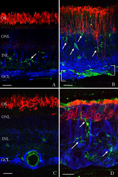
Figure 5. Localization of microglia in normal and detached human retina
Laser scanning confocal images are shown of human retina labeled with the lectin Ricinus communis (green) and antibodies to rod opsin (red) and glial fibrillary acidic protein (GFAP; blue). A: In normal retina the lectin labels cells in the inner plexiform layer; anti-rod opsin is present in the rod outer segments (OS) and anti-GFAP is present in Müller cells and astrocytes. B: In longstanding detachments (exact duration unknown but at least 1 month), lectin-labeled cells (arrows) are dispersed throughout the retina and within an epiretinal membrane (brackets). Anti-opsin labeling is elevated in the outer nuclear layer (ONL) and rod axons are observed extending into the inner retina. Anti-GFAP labeling of Müller cells is elevated. C: An attached retina from an eye with an exudative detachment as a result of a malignant melanoma is shown illustrating the normal distribution of lectin, anti-opsin, and anti-GFAP labeling. The large lectin-labeled structure in the ganglion cell layer (GCL) is a blood vessel. D: A detached retina is shown from the same eye as shown in C illustrating the numerous lectin-labeled cells dispersed throughout the retina (arrows), elevated anti-opsin labeling in the ONL, and anti-GFAP labeling of Müller cells. The inner nuclear layer (INL) is also identified. Scale bars represent 20 μm.