![]() Figure 4 of
Lewis, Mol Vis 2005;
11:491-500.
Figure 4 of
Lewis, Mol Vis 2005;
11:491-500.
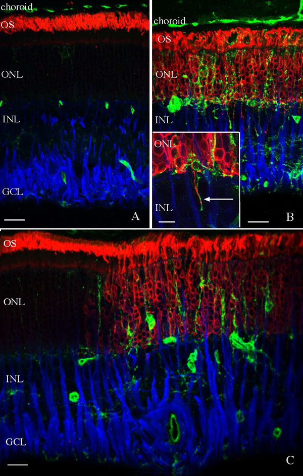
Figure 4. Localization of microglia in the reattached feline retina
Laser scanning confocal images are shown of feline retina, detached for 3 days and reattached for 28 days, labeled with the lectin Griffonia Simplicifolia (green) and antibodies to rod opsin (red) and glial fibrillary acidic protein (GFAP; blue). A: A typical region of reattached retina demonstrates that few lectin labeled cells are present in the outer nuclear layer (ONL) at this time. Anti-rod opsin labeling is present in the now organized outer segments (OS) and anti-GFAP labeling extends into the inner nuclear layer (INL). B: In focal "patchy" regions with poor photoreceptor recovery, as illustrated by the continued anti-opsin labeling of the ONL and disorganized OS, many lectin labeled cells are present throughout the retina. Lectin-labeled processes are often observed in close association with rod axons that extend into the inner retina following reattachment (arrow). C: A reattached retina is shown illustrating the abrupt transition from retinal areas with few lectin labeled cells in the ONL (left half of image) to regions with many lectin-labeled cells in the ONL (right half of image). The lectin-labeled cells present in the ONL occur predominantly in regions of poor photoreceptor recovery as illustrated by the increased anti-opsin labeling. The lectin also labeled blood vessels in the retina and choroid. The ganglion cell layer (GCL) is also identified. Scale bars represent 20 μm in A,B,C and 10 μm in the inset to B.