![]() Figure 3 of
Lewis, Mol Vis 2005;
11:491-500.
Figure 3 of
Lewis, Mol Vis 2005;
11:491-500.
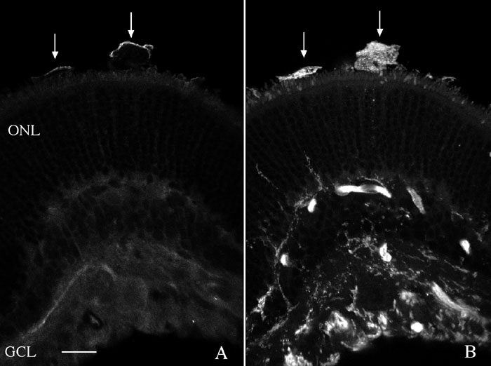
Figure 3. Localization of Griffonia simplicifolia and CD11b in the feline retina
Laser scanning confocal images are shown of a 3-day detached feline retina labeled with anti-CD11b (A) and the lectin Griffonia simplicifolia (B). A single section was originally double labeled but the two channels are separated here to more easily visualize the labeling patterns. A: Anti-CD11b labeling is present only in the presumptive macrophages in the subretinal space (arrows). B: The same section shown in A demonstrating that the lectin labels cells throughout the retina, in the subretinal space (arrows), and blood vessels. The outer nuclear layer (ONL) and ganglion cell layer (GCL) are also identified. The scale bar represents 20 μm.