![]() Figure 2 of
Lewis, Mol Vis 2005;
11:491-500.
Figure 2 of
Lewis, Mol Vis 2005;
11:491-500.
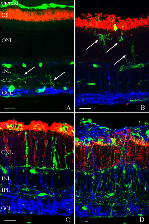
Figure 2. Localization of microglia in the normal and detached feline retina
Laser scanning confocal images of feline retina are shown labeled with the lectin Griffonia simplicifolia (green) and antibodies to rod opsin (red) and glial fibrillary acidic protein (GFAP; blue). A: In normal retina, the lectin labels microglia in the inner plexiform layer (IPL; arrows) and blood vessels in the retina and choroid. Anti-rod opsin labels rod outer segments (OS). Anti-GFAP labels Müller cell endfeet and astrocyte processes. B: One day after retinal detachment, lectin labeled cells are observed in the outer nuclear layer (ONL) as rod OS begin to degenerate and Müller cells upregulate GFAP expression. C: Three days after retinal detachment lectin-labeled cells with varied morphologies are distributed throughout the inner and outer retina and in the OS layer. Anti-rod opsin is present in the degenerating OS and in the plasma membrane of some rod photoreceptors. In the ONL and anti-GFAP, labeling extends throughout the retina. D: Twenty-eight days after retinal detachment, numerous lectin-labeled cells are present throughout the retina and in a subretinal glial scar labeled with anti-GFAP. Anti-rod opsin labeling illustrates the extensive OS degeneration and redistribution of the protein to the entire photoreceptor plasma membrane at this time. The inner nuclear layer (INL) and ganglion cell layer (GCL) are also identified. The scale bars represent 20 μm.