![]() Figure 1 of
Lewis, Mol Vis 2005;
11:491-500.
Figure 1 of
Lewis, Mol Vis 2005;
11:491-500.
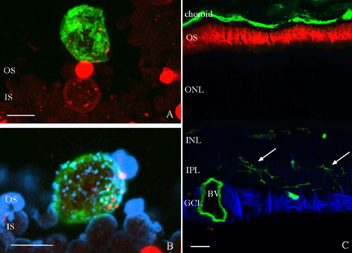
Figure 1. Lectin labeling of detached and attached feline retina
A: A laser scanning confocal image is shown of a cell in the subretinal space from 3 day detached feline retina labeled with the lectin Griffonia simplicifolia (green) and antibodies to M cone opsin (red). The cone outer segment (OS) is truncated and the opsin is redistributed to the inner segment (IS). Cone opsin material is present within the cell (small dots). B: A laser scanning confocal image is shown of a cell in the subretinal space from 3 day detached feline retina labeled with the lectin Griffonia simplicifolia (green) and antibodies to both rod opsin (blue) and M cone opsin (red). The cell (green) appears in association with the degenerate rods but contains both rod and cone opsin labeled material (small dots). C: A laser scanning confocal image is shown of attached feline retina from an eye with a 28 day detachment, labeled with the lectin Griffonia simplicifolia (green) and antibodies to rod opsin (red) and glial fibrillary acidic protein (GFAP; blue). No significant activation of microglia is apparent as microglia continue to reside in the inner plexiform layer (IPL; arrows). The outer segments (OS), outer nuclear layer (ONL), inner nuclear layer (INL), ganglion cell layer (GCL), and blood vessels (BV) are also identified. The scale bars represent 5 μm in A,B and 20 μm in C.