![]() Figure 2 of
Roldan-Pallares, Mol Vis 2005;
11:461-471.
Figure 2 of
Roldan-Pallares, Mol Vis 2005;
11:461-471.
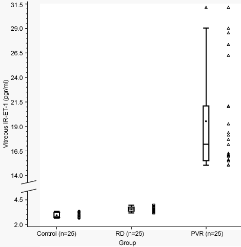
Figure 2. Vitreous IR-ET-1 levels
Vitreous IR-ET-1 levels (pg/ml) of the three groups of patients represented as box and whisker plots (explained in Figure 1). The bottom and top edges of the box are located at the 25th and 75th percentiles of the sample. The dot inside the box represents the mean. The median (50th percentile) is shown as a horizontal line in the box. The vertical lines extend from the box as far as the data extend, to a distance of at most 1.5 interquantile ranges. Any value more extreme than this is marked with a plot symbol.