![]() Figure 6 of
Tasheva, Mol Vis 2005;
11:452-460.
Figure 6 of
Tasheva, Mol Vis 2005;
11:452-460.
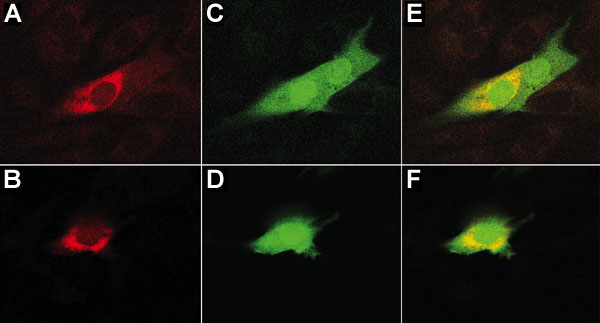
Figure 6. Colocalization of B7 and mimecan proteins
The subcellular localization of B7 and mimecan fusion proteins was determined using transient transfections and confocal microscopy. MG-63 cells were co-transfected with pEGFP.B7 and pcDNA-Exp40/Bovine mimecan constructs. After transfection, the cells were incubated with primary mouse anti-V5 antibody followed by incubation with Alexa Fluor 546 conjugated secondary antibody and examined by confocal microscopy. Two examples each of mimecan staining (A,B) and B7 staining (C,D) are shown. Confocal microscopy images (E,F) show the merger of the adjacent pairs of images.