![]() Figure 3 of
Tasheva, Mol Vis 2005;
11:452-460.
Figure 3 of
Tasheva, Mol Vis 2005;
11:452-460.
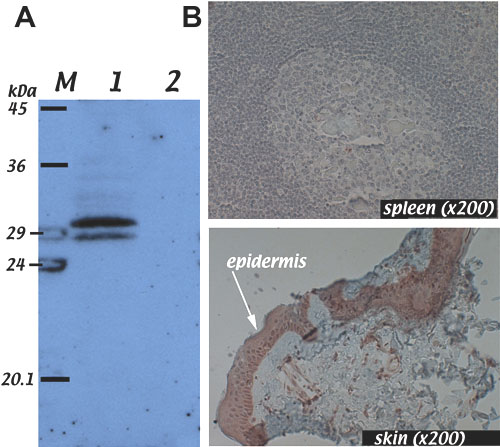
Figure 3. Specificity of B7.N antibody
Western blot analysis was performed to test whether the B7.N antibody recognized isoform 2 of B7. IHC analysis was performed to confirm the specificity of our antibody. A: In lane 1, the antibody B7.N selectively recognizes the 31 kDa isoform 2 of B7 that was transcribed and translated in vitro from the pReceiver.B7 construct. Lane 2 shows an immunoblot from an in vitro transcription/translation reaction that was performed without addition of pReceiver.B7 template. B: Immunohistochemistry shows the presence of B7 in the epidermal layer of the skin (arrow) and its absence in spleen. Positive immunostaining appears brown.