![]() Figure 5 of
Fang, Mol Vis 2005;
11:443-451.
Figure 5 of
Fang, Mol Vis 2005;
11:443-451.
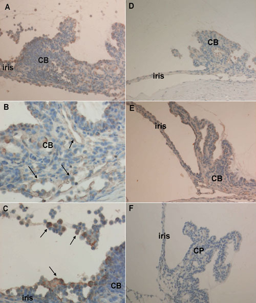
Figure 5. Immunohistochemical staining for fractalkine expression in the iris/ciliary body from Lewis rats
Rats with experimental autoimmune anterior uvietis showed stronger fractalkine expression in vascular endothelial cells (arrows) in the ciliary body (CB; A,B) and in the infiltrating cells (arrows) in the iris/CB and posterior chamber (A,C), compared with those of normal rats (E). Treatment with PDTC (200 mg/kg/day) contributed to reduced mononuclear cell infiltration and fractalkine expression in the iris/CB (D). The negative control (F) was obtained using goat IgG as primary antibody. A ciliary process (CP) is also labeled. The original magnifications were 200x in A,D,E and 400x in B,C.