![]() Figure 1 of
Tsai, Mol Vis 2005;
11:50-55.
Figure 1 of
Tsai, Mol Vis 2005;
11:50-55.
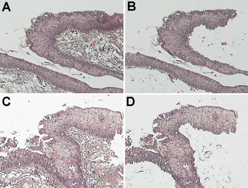
Figure 1. Representative results of laser capture micro-dissection (LCM) of pterygium
A,C: The epithelium and subepithelial fibrovascular layer of two specimens before micro-dissection. B,D: Only epithelium remains in specimens after micro-dissection. The images in A and B are from one specimen; the images in C and D are from another specimen.