![]() Figure 1 of
Challa, Mol Vis 2005;
11:425-430.
Figure 1 of
Challa, Mol Vis 2005;
11:425-430.
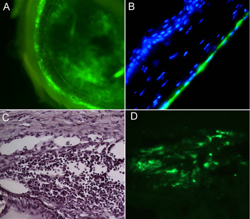
Figure 1. GFP expression in the anterior segments of rodents transfected with a lentiviral vector
Lentiviral vectors were constructed using the EF-1α promoter to drive expression of enhanced green fluorescent protein (GFP). After intracameral injection of the viral vector, successful transfection is seen in the trabecular meshwork (TM), Schlemm's canal, and corneal endothelium. A: GFP expression in a globe whole mount. The most intense expression is seen in regions of the TM followed by lower expression in the cornea. B: A section of the cornea, counterstained with DAPI, in which GFP expression is seen in the corneal endothelium. C: Hematoxylin staining of the anterior chamber angle. The TM and Schlemm's canal are seen. D: Intense GFP expression in the cells of the TM.