![]() Figure 2 of
Hollborn, Mol Vis 2005;
11:397-413.
Figure 2 of
Hollborn, Mol Vis 2005;
11:397-413.
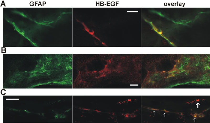
Figure 2. HB-EGF and GFAP immunoreactivity in fibroproliferative PVR membranes
HB-EGF and GFAP immunoreactivity in surgically excised fibroproliferative membranes from three patients with PVR. The epiretinal membranes were double stained with antisera against the glial cell marker GFAP (green fluorescence) and HB-EGF (red fluorescence). Coexpression of both proteins yields a yellow staining (overlay). A: In a PVR membrane, there were GFAP expressing cells that showed no HB-EGF immunoreactivity and glial cell bodies that were double stained against HB-EGF. B: A similar pattern was observed in another PVR membrane, with GFAP expressing structures with or without HB-EGF colabeling. C: In epiretinal PVR membranes, the predominant GFAP expressing structures were long and thin processes. These fibrillary processes were partially immunoreactive for HB-EGF protein (small arrows) and partially devoid of HB-EGF immunoreactivity. Additionally, there were HB-EGF expressing structures without double labeling with GFAP (large arrow). Similar staining patterns were obtained in six different membranes. Scale bars represent 20 μm.