![]() Figure 1 of
Chidlow, Mol Vis 2005;
11:387-396.
Figure 1 of
Chidlow, Mol Vis 2005;
11:387-396.
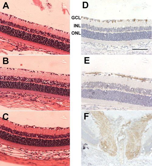
Figure 1. Effect of ONT on retinal histology and NF-L immunohistochemistry
A-C: Light photomicrographs from paraffin embedded transverse sections of normal and axotomized rat retinas stained with hematoxylin and eosin. A: Retina from control untreated eye. B: Retina from vehicle injected eye 7 days after ONT. C: Retina from BDNF (5 μg) injected eye 7 days after ONT. D-F: Light photomicrographs of NF-L immunohistochemistry in normal rat retina and optic nerve head. D: NF-L labeling is associated with RGC soma in the GCL but not with soma in the INL or ONL. E: NF-L labeling is associated with RGC axons. F: NF-L labeling is associated with axons in the optic nerve head. Thus, NF-L is associated with RGCs but not with other cell types in the rat retina.