![]() Figure 3 of
Ogawa, Mol Vis 2005;
11:380-386.
Figure 3 of
Ogawa, Mol Vis 2005;
11:380-386.
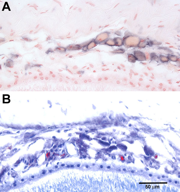
Figure 3. Staining of macrophages and mast cells in the choroid
A: Macrophage cell staining (blue-purple color) using a rat anti-mouse F4/80 antigen monoclonal antibody on eye tissue from a 21-month-old male C57BL/6 mouse. B: Mast cell staining (red) using toluidine blue O (0.1%) on eye tissue from a 21-month-old male C57BL/6 mouse. Differential staining was enhanced using Photoshop. The original magnification in both images was 460x. The scale bar represents 50 μm.