![]() Figure 1 of
Ogawa, Mol Vis 2005;
11:380-386.
Figure 1 of
Ogawa, Mol Vis 2005;
11:380-386.
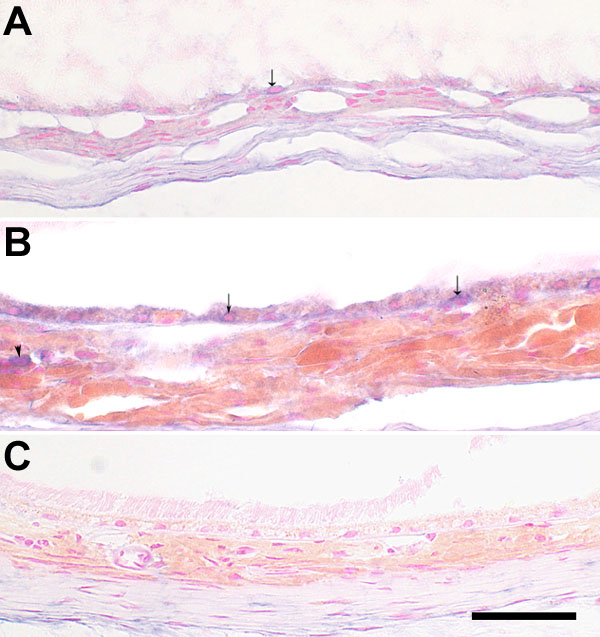
Figure 1. Labeling of cathepsin S mRNA by in situ hybridization in the RPE/choroid
A: RPE/choroid sections from a 2-month-old C57BL/6 mouse labeled with antisense RNA. B: RPE/choroid sections from a 24-month-old C57BL/6 mouse labeled with antisense RNA. C: RPE/choroid sections from a 24-month-old C57BL/6 mouse labeled with sense RNA probe. The digoxigenin labeled in situ hybridization reaction with BCIP/NBT appears blue-purple. Arrows show cathepsin S (CatS) labeled RPE cells. CatS labeled choroidal cells (arrowhead) were seen in the 2-month-old sections (A) and 24-month-old sections (B). Only a few representative positive cells are marked with arrows or arrowheads. No labeled cells were observed in the RPE and choroid with sense RNA probe (C). The scale bar represents 50 μm.