![]() Figure 3 of
Kinose, Mol Vis 2005;
11:366-373.
Figure 3 of
Kinose, Mol Vis 2005;
11:366-373.
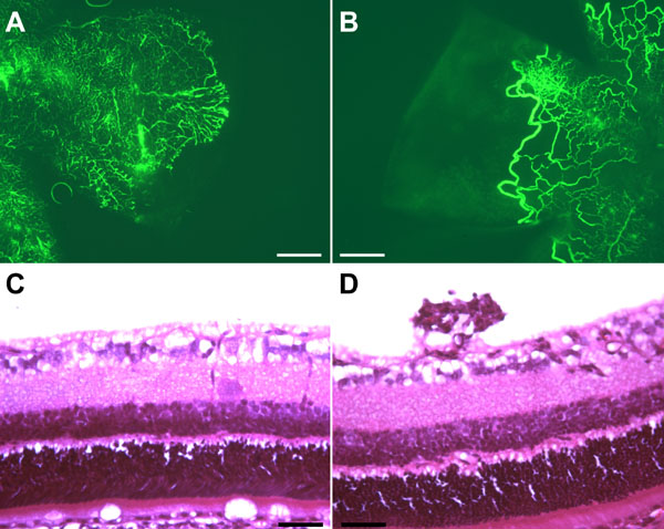
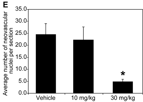
Figure 3. Oral dosing of Compound A decreases neovascularization in a rat OIR model
A,B: Representative 100x images of retinal flat mounts from postnatal day 21 (P21) rat pups treated with alternating cycles of 50% and 10% oxygen for 14 days, followed by oral vehicle (A) or 30 mg/kg kinase inhibitor (B) dosing for 7 days. The vasculature was visualized by fluorescence microscopy of P21 fluorescein-dextran perfused retinas dissected and mounted on microscope slides. C,D: Representative hematoxylin and eosin stained sections from 6 μm sections of P21 rat retinas showing the ganglion cell/vitreous interface. C: Retina from a rat raised in room air from P0-P21. D: Retina from a rat raised in cycling oxygen for 14 days, followed by 7 days in room air. Note the presence of preretinal (above the ganglion cell layer) vessels in the oxygen reared rat (D) and their absence in the room air reared rat (C). E: Quantitative analysis of the number of preretinal neovascular nuclei per retinal section from the various treatment groups. An average of 180 sections through each eye of vehicle treated (n=16), 10 mg/kg kinase inhibitor treated (n=8), and 30 mg/kg kinase inhibitor treated (n=14) rats were counted. Results are presented as the average number of preretinal nuclei/section per animal. Error bars represent standard errors of the mean. Asterisk indicates t-test p value<0.005 as compared to vehicle treatment. Scale bar represents 200 μm in panels A and B and 50 μm in panels C and D.