![]() Figure 2 of
Kinose, Mol Vis 2005;
11:366-373.
Figure 2 of
Kinose, Mol Vis 2005;
11:366-373.
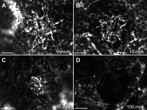
Figure 2. Oral dosing of Compound A potently inhibits neovascular lesion size in a rat laser CNV model
Oral dosing of Compound A inhibits neovascular lesion size in a rat laser CNV model in a dose-dependent manner. Representative lesions from choroidal flat-mounts of fluorescein-dextran perfused adult male Brown Norway rats dosed with 0 mg/kg (A), 10 mg/kg (B), 30 mg/kg (C), or 100 mg/kg (D) Compound A. Prior to fluorescein-dextran perfusion, small breaks in Bruch's membrane were made using an argon blue-green ophthalmic laser, and the animals were treated with the indicated dose of kinase inhibitor by oral gavage for 12 days. The neovascular lesions were visualized at 200x magnification using a fluorescence microscope. Images of fluorescent vessels in the lesions were captured and quantitated using ImagePro Plus. E: Quantitative assessment of neovascular lesion size. The average lesion size from each animal was calculated using ImagePro Plus and plotted against compound dose (6 per dosing group). Asterisk indicates t-test p value<0.0001 compared to vehicle treatment. Scale bar represents 100 μm.