![]() Figure 3 of
Salvador-Silva, Mol Vis 2005;
11:356-365.
Figure 3 of
Salvador-Silva, Mol Vis 2005;
11:356-365.
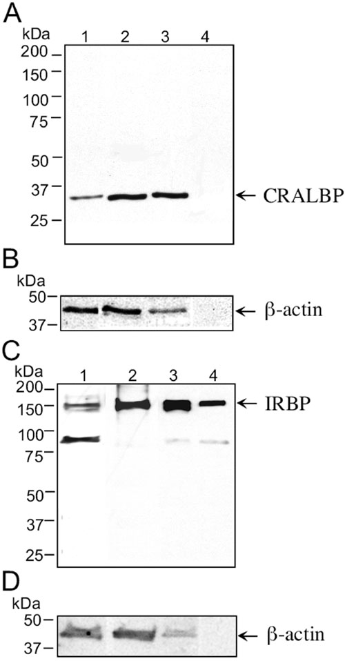
Figure 3. Western blot patterns of CRALBP and IRBP in the pars plicata region of the ciliary epithelium
Whole tissue extracts (30 μg/lane) from the bovine ciliary processes (lane 1), RPE (lanes 2), retina (lane 3), and aqueous humor (lanes 4) were resolved by 4-15% SDS-PAGE gels and subjected to immunoblot analysis using a polyclonal antibody against CRALBP (A), a monoclonal antibody against bovine IRBP (C) and a β-actin antibody (B,D). The antigen-antibody complexes were detected by the ECL chemiluminescent system. The migration of CRALBP (36 kDa) and IRBP (140 kDa) are marked with arrows.