![]() Figure 2 of
Salvador-Silva, Mol Vis 2005;
11:356-365.
Figure 2 of
Salvador-Silva, Mol Vis 2005;
11:356-365.
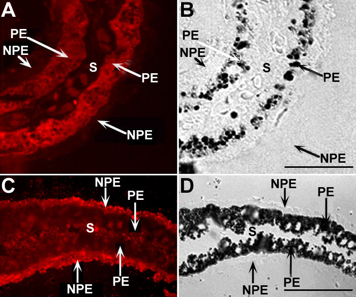
Figure 2. Cellular distribution of CRALBP and IRBP on semithin cryostat sections of the bovine ciliary epithelium
Cryostat sections from the pars plicata region were labeled with a polyclonal antibody against CRALBP (A,B) or a monoclonal antibody to IRBP (C,D). Photographs of B and D are the phase contrast of the indirect immunofluorescence photographs of A and C, respectively. The nonpigmented epithelium (NPE), pigmented epithelium (PE), and stroma (S) are labeled. Scale bar represents to 1 μm.