![]() Figure 5 of
Reiners, Mol Vis 2005;
11:347-355.
Figure 5 of
Reiners, Mol Vis 2005;
11:347-355.
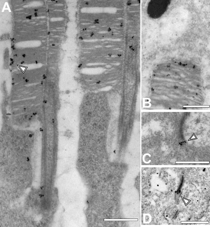
Figure 5. Immunoelectron analysis of Pcdh15 in photoreceptor cells
Silver enhanced immunogold labeling of Pcdh15 in ultrathin sections through parts of mouse photoreceptor cells. A: Longitudinal section through a portion of outer segments (OS) and apical inner segments of rod photoreceptor cells. Silver enhanced immunogold particles were associated with OS disks and accumulated at the OS plasma membrane (arrowhead). B: Section through the apical part of a RPE cell and the tip of an OS. Silver enhanced immunogold labeling was restricted to the OS. A RPE cells the labeling was not above the background. C,D: A section through synaptic terminals of photoreceptor cells. Silver enhanced immunogold labeling of Pcdh15 (C) and harmonin (D) was present in the ribbons of photoreceptor synapses (arrowheads). The scale bars represent 0.5 μm.