![]() Figure 3 of
Reiners, Mol Vis 2005;
11:347-355.
Figure 3 of
Reiners, Mol Vis 2005;
11:347-355.
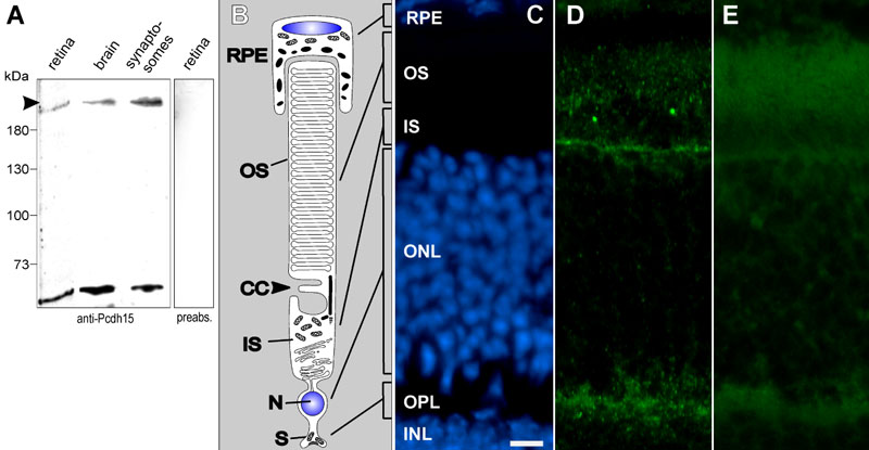
Figure 3. Pcdh15 expression in the retina
A: Immunoblot of total protein lysates of adult murine retina, brain, and brain synaptosomes with anti-Pcdh15. In lanes 1-3 two bands were obtained, one at the estimated molecular mass of holo-Pcdh15 (214 kDa, arrow) and one at about 65 kDa representing the Pcdh15 isoform b. Both bands were abolished by preabsorption of anti-Pcdh15 with recombinant Pcdh15 (lane 4). B: Scheme of a rod photoreceptor cell. Vertebrate photoreceptors are composed of a light sensitive outer segment (OS) linked via a connecting cilium (CC) to an inner segment (IS) which contains the biosynthetic and metabolic machinery. The nucleus (N) is localized in the outer nuclear layer (ONL) and S the synaptic terminal (S) is located in the outer plexiform layer (OPL) of the retina. C,E: Fluorescence microscopy of a longitudinal cryosection through a mouse retina. C: DAPI staining [8] present in the nuclei of the RPE [9] cells, ONL, and the inner nuclear layer (INL) separated by the OPL. D: Indirect immunofluorescence of anti-Pcdh15 was found in photoreceptor synapses within OPL and the photoreceptor outer segments with occasional intense dot-like structures at the outer segment base. Furthermore, the outer limiting membrane just proximal to the ONL was stained by anti-Pcdh15. The scale bar represents 5 μm. E: In the absence of primary antibodies, no fluorescence is detected.