![]() Figure 2 of
Reiners, Mol Vis 2005;
11:347-355.
Figure 2 of
Reiners, Mol Vis 2005;
11:347-355.
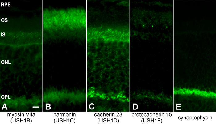
Figure 2. Colocalization of USH1 gene products at photoreceptor synapses
Indirect immunofluorescence of antibodies against myosin VIIa (A), harmonin (B), Cdh23 (C), Pcdh15 (D), and synaptophysin (E) in parallel longitudinal cryosections through a mouse retina. Anti-myosin VIIa (A) labeling was present in retinal pigmented epithelium (RPE) cells, in connecting cilia at junctions between outer segments (OS) and inner segments (IS) and at photoreceptor synapses in the outer plexiform layer (OPL). Harmonin (B) was localized in the OS, IS and OPL. Cdh23 (C) was expressed in the IS and OPL. As shown in previous figures, Pcdh15 (D) was found in the OS, predominantly at its base, and in the OPL. All four USH1 proteins colocalize in the OPL where ribbon synapses of photoreceptor cells are localized. The synaptic region is visualized by the synaptic marker anti-synaptophysin (E). Harmonin, Cdh23, and Pcdh15 are not expressed in the RPE. The outer nuclear layer (ONL) is also labeled. The scale bar represents 5 μm.