![]() Figure 4 of
Paull, Mol Vis 2005;
11:328-334.
Figure 4 of
Paull, Mol Vis 2005;
11:328-334.
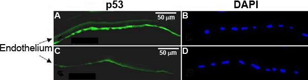
Figure 4. Expression of p53 in the corneal endothelium
A,B: Central cornea. C,D: Peripheral cornea. Fluorescence for p53 is shown in green (A,C) and DAPI fluorescence is shown in blue (B,D). DAPI stains DNA and confirms that cells were present in both central and peripheral sections. These results show stronger staining for p53 in the central endothelium compared to the peripheral endothelium. All micrographs are representative of multiple sections of the cornea. The brightness, contrast, and exposure times were not adjusted between micrographs of central and peripheral tissue.